Book a visit
+371 26 412 412
We Treat
We help you regain your health and quality of life by treating various gynaecological conditions. Our experienced specialists provide a personalised approach, guided by the latest medical standards and gentle treatment methods.

Diastasis

What is diastasis?

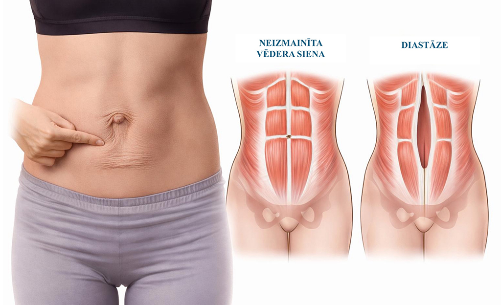
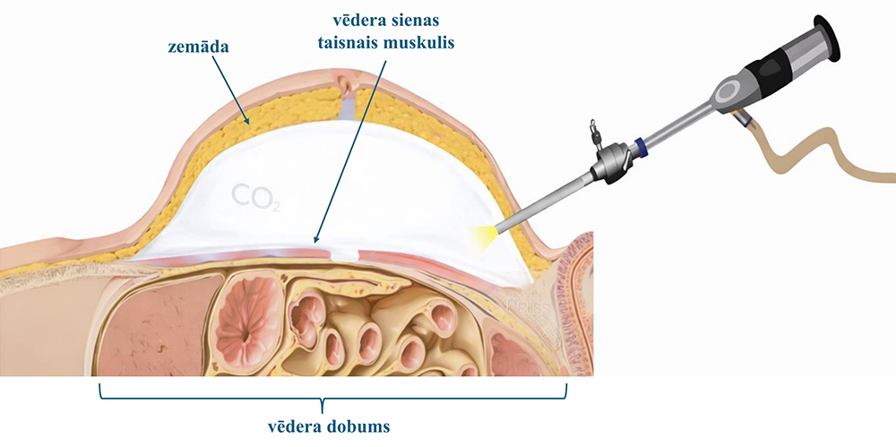
Diastasis is a condition in which a gap forms along the abdominal midline between the rectus muscles of the anterior abdominal wall, causing abdominal wall weakness (Figure 1). Diastasis is not a hernia, because there is no fascial defect (in the connective tissue layer of the abdominal wall), although both conditions may coexist.


Vēdera priekšējās sienas diastāze
Figure 1. Diastasis of the anterior abdominal wall

Why does diastasis develop?

Diastasis is promoted by several factors: pregnancy, obesity, heavy physical exertion (incorrect technique) and ageing (weakening of connective tissue).

How is diastasis diagnosed?

Diastasis is diagnosed clinically when the physician assesses the anterior abdominal wall. It can also be identified by self-examination, following the steps below:

  1. Lie on your back: knees bent, feet on the floor, abdomen relaxed.
  2. Place 2-3 fingers on the abdominal midline. Start at the umbilical region, then assess above and below the navel (up to the palpable bony structures: the xiphoid process superiorly and the pubic bone inferiorly).
  3. Gently lift the head and shoulders, feeling the abdominal wall muscles tense.
  4. Assess the width of the gap between the muscles in finger widths (turn the fingers perpendicular to measure the width).
  5. A distance of 0-2 finger widths is an acceptable finding; 2-3 finger widths indicate mild diastasis; >3 finger widths indicate marked diastasis.

For more precise assessment and measurement of diastasis, ultrasonography (US) is used, usually ordered by a specialist.


What are the symptoms of diastasis?

Complaints are most often cosmetic and functional. A dome-shaped protrusion appears in the abdominal midline during exertion or changes in body position, and persistent body-contour changes may develop. Because abdominal wall weakness affects core stability, more pronounced diastasis may be accompanied by postural disorders, low back pain, pelvic floor muscle dysfunction (pelvic heaviness, urinary incontinence), increased bloating after meals and difficulty with daily physical activities. Diastasis also increases the risk of an abdominal wall hernia.

What is the treatment for diastasis?

Treatment of diastasis is gradual and depends on symptoms, diastasis width and the patient's goals. The two main approaches are conservative therapy (physiotherapy) and surgical treatment.

Surgical treatment is indicated in cases where:

  • there is marked diastasis (greater than 5cm);
  • diastasis is at least 3cm and is accompanied by other symptoms;
  • functional symptoms related to diastasis are observed (pain, core instability);
  • physiotherapy is ineffective;
  • diastasis exists in combination with a hernia.

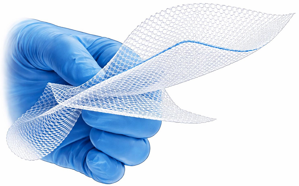
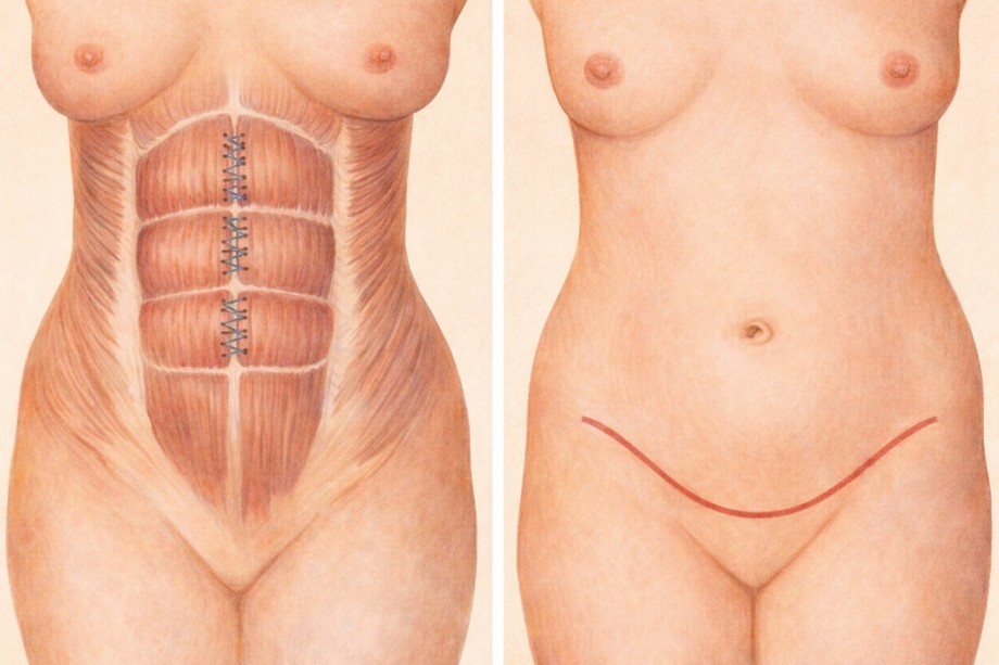
The main surgical methods for diastasis correction are laparoscopic and conventional (open) approaches. The aim is to suture the edges of the rectus muscles of the anterior abdominal wall together while correcting an abdominal wall hernia if present. Technique choice depends on abdominal wall tissue quality, diastasis width and coexisting hernias. Usually, a synthetic mesh is inserted to strengthen the abdominal wall and reduce the risk of diastasis recurrence (reformation) (Figure 2).

Figure 3. Synthetic mesh
Figure 2. Synthetic mesh

Minimally invasive correction of diastasis is performed mainly using one of the following techniques:

  • TAPP (Transabdominal Preperitoneal Approach) – laparoscopic approach (entering the abdominal cavity) (Figure 3);
Vēdera dobums
Figure 3. TAPP technique
  • eTEP (Enhanced-View Totally Extraperitoneal repair) endoscopic extraperitoneal approach (without entering the abdominal cavity), in which a space is dissected between the posterior sheath of the rectus abdominis muscle and the muscle itself (Figure 4);
Vēdera dobums
Figure 4. eTEP technique
  • REPA (Endoscopic Pre-Aponeurotic Repair) / SCOLA (Subcutaneous Onlay Laparoscopic Approach) – endoscopic preaponeurotic approach (without entering the abdominal cavity), in which a space is dissected between the anterior sheath of the rectus abdominis muscle and the subcutaneous tissue (Figure 5).
Vēdera dobums
Figure 5. REPA / SCOLA technique

Conventional or open surgery for correction of diastasis (if the only aim is to eliminate the abdominal wall defect) is performed much less often, because minimally invasive approaches are the current standard of care for diastasis. If excess abdominal skin must also be removed (abdominoplasty), correction of diastasis and hernias can be performed at the same time (Figure 6).

Konvencionāla diastāzes korekcija
Figure 6. Conventional correction of diastasis combined with abdominoplasty


Doctors to See


Working hours
Weekdays
8.00 - 19.00
Weekends
By appointment
Address
Medical centre New Hanza Med
Mihaila Tāla iela 1, Floor 4–6
Riga, Latvia
Contacts
info@vitolaklinika.lv
+371 26 412 412
+371 27 835 618
In partnership with:
Compensa_logotipsSeesam_logo
© 2026 Vītola klīnika
All rights reserved