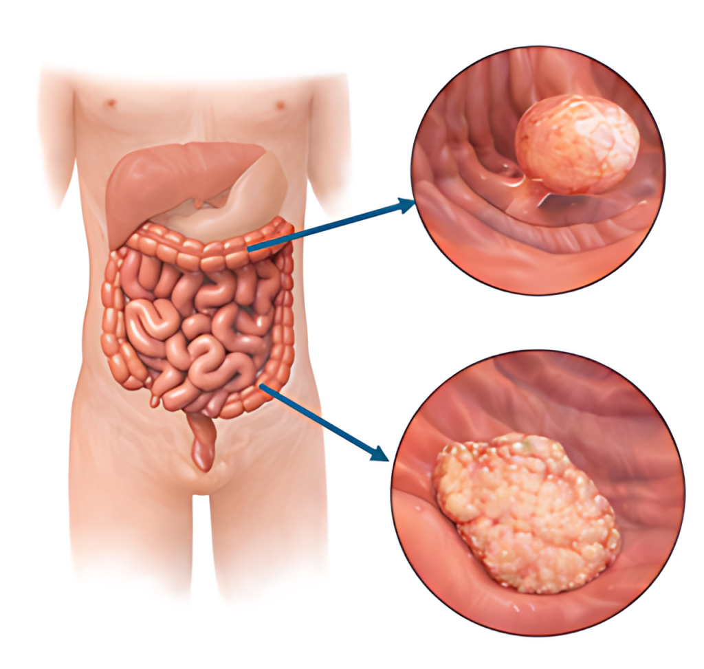
Colonic polyps are slowly growing mucosal lesions (pedunculated or flat) that are very common in the general population, and their prevalence increases with age (Figure 1). Depending on their subtype, polyps have a higher or lower risk of malignant transformation; therefore, they must always be removed when detected.
The development of colonic polyps is influenced by several factors – genetic mutations in cells of the intestinal wall (occurring during life), ageing, diet (a low-fibre diet and high consumption of processed food and red meat), harmful habits (smoking, alcohol use), a sedentary lifestyle, obesity, chronic inflammation in the colon, inherited genetic syndromes.
In most patients, colonic polyps do not cause complaints; likewise, their presence cannot be determined by laboratory tests. For this reason, colonoscopy is the only way to diagnose them in a timely manner. Starting from 50 years of age (in some countries – starting from 45 years), colonoscopy is recommended; it is both a diagnostic and therapeutic procedure (allows polyps to be detected and removed). The age at which colonoscopy is performed and the frequency of the examination may vary depending on individual risk factors, for which consultation with a physician is necessary.
Colonic polyps mostly do not cause symptoms. If they appear, they are most likely already associated with a large polyp or its malignant transformation. Colonic polyps or colon cancer may be indicated by – bleeding from the rectum, anaemia, changes in bowel movement habits.
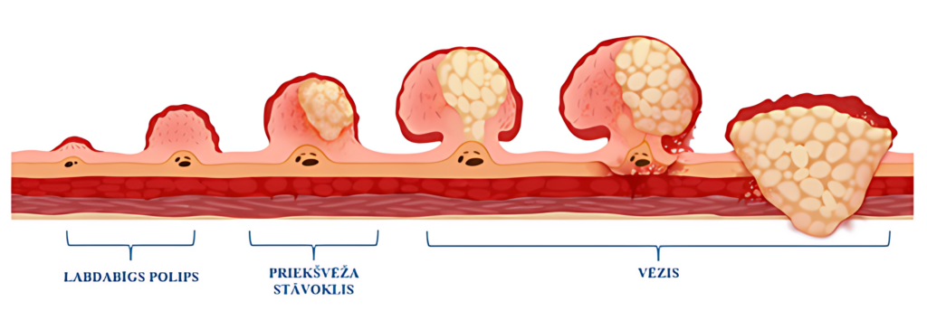
Untreated colonic polyps may progress over several years to a malignant tumour, which most often occurs unnoticed, without pronounced symptoms (Figure 2). Larger polyps may also cause chronic bleeding that is not noticeable during bowel movements but is reflected in laboratory analyses – as a positive faecal occult blood test and chronic anaemia in blood tests.
If a small polyp is detected during colonoscopy, it is removed already during the diagnostic procedure. If the size, location, number or other visual characteristics of the polyp do not allow endoscopic polypectomy, a biopsy is performed to assess its microscopic structure (histological examination), and surgical treatment should be planned.
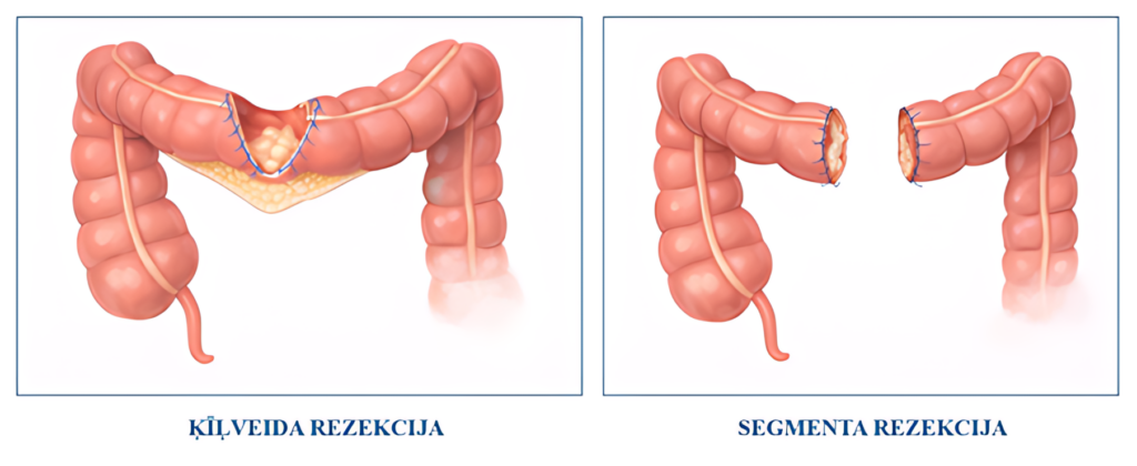
The extent of surgical treatment differs depending on the size of the polyp, anatomical location and histological examination result. The operation is performed laparoscopically, and during it wedge excision of the colon (excision of part of one intestinal wall, including the polyp) or segmental resection (complete division of the intestine and removal of a small segment together with the polyp) is performed (Figure 3).
Most often, before this it is necessary to mark the polyp (during colonoscopy), so that the location of the lesion can be visualised laparoscopically and surgical treatment can be as sparing as possible.


CALL US:
+371 26 412 412WHATSAPP:
+371 26 412 412
If you need an appointment before surgery, please let us know when calling —
we will offer you the earliest available appointment.
SEND US A MESSAGE